Copyright
©The Author(s) 2016.
World J Radiol. Aug 28, 2016; 8(8): 757-763
Published online Aug 28, 2016. doi: 10.4329/wjr.v8.i8.757
Published online Aug 28, 2016. doi: 10.4329/wjr.v8.i8.757
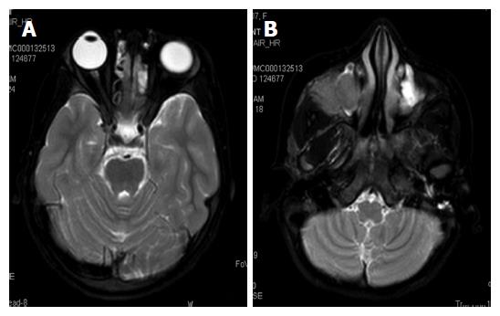
Figure 6 The whole body magnetic resonance imaging showing presence of bilateral ethmoid and maxillary sinusitis along with replacement of normal marrow by focal lesions involving maxilla.
A: Axial TSE T2-weighted fat suppressed image of normal orbits; B: T2WFS axial image of the face showing isointense soft tissue filling the right maxillary sinus with extension into the infratemporal fossa. TSE: Turbo spin-echo; T2WFS: T2-weighted fat-suppressed.
- Citation: Vallonthaiel AG, Mridha AR, Gamanagatti S, Jana M, Sharma MC, Khan SA, Bakhshi S. Unusual presentation of Erdheim-Chester disease in a child with acute lymphoblastic leukemia. World J Radiol 2016; 8(8): 757-763
- URL: https://www.wjgnet.com/1949-8470/full/v8/i8/757.htm
- DOI: https://dx.doi.org/10.4329/wjr.v8.i8.757


