Copyright
©The Author(s) 2016.
World J Radiol. Aug 28, 2016; 8(8): 757-763
Published online Aug 28, 2016. doi: 10.4329/wjr.v8.i8.757
Published online Aug 28, 2016. doi: 10.4329/wjr.v8.i8.757
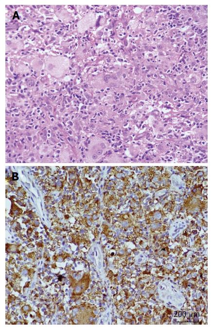
Figure 3 A biopsy from the lesion.
A: Hematoxylin and eosin stained section showing an infiltrate comprised of foamy and lipid laden histiocytes, multinucleated giant cells, lymphocytes and fibroblastic cells (200 ×); B: Immunohistochemistry with CD68 showing positivity in histiocytic cells and giant cells (200 ×).
- Citation: Vallonthaiel AG, Mridha AR, Gamanagatti S, Jana M, Sharma MC, Khan SA, Bakhshi S. Unusual presentation of Erdheim-Chester disease in a child with acute lymphoblastic leukemia. World J Radiol 2016; 8(8): 757-763
- URL: https://www.wjgnet.com/1949-8470/full/v8/i8/757.htm
- DOI: https://dx.doi.org/10.4329/wjr.v8.i8.757


