Copyright
©The Author(s) 2016.
World J Radiol. Aug 28, 2016; 8(8): 757-763
Published online Aug 28, 2016. doi: 10.4329/wjr.v8.i8.757
Published online Aug 28, 2016. doi: 10.4329/wjr.v8.i8.757
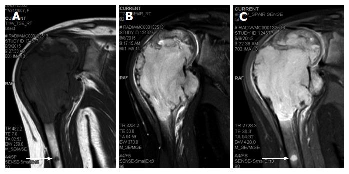
Figure 2 Oblique coronal turbo spin-echo T1-weighted (A), turbo spin-echo T2-weighted fat-suppressed (B) and proton-density-weighted fat-suppressed (C) magnetic resonance imaging images of the right shoulder showing a solid expansile intramedullary mass replacing the normal marrow fat; hypointense on T1-weighted images, hyperintense on T2-weighted images.
A cortical break in medial upper humeral diaphysis with extension into soft tissue noted. Note another skip lesion in the humeral shaft with similar signal characteristics (white arrow).
- Citation: Vallonthaiel AG, Mridha AR, Gamanagatti S, Jana M, Sharma MC, Khan SA, Bakhshi S. Unusual presentation of Erdheim-Chester disease in a child with acute lymphoblastic leukemia. World J Radiol 2016; 8(8): 757-763
- URL: https://www.wjgnet.com/1949-8470/full/v8/i8/757.htm
- DOI: https://dx.doi.org/10.4329/wjr.v8.i8.757


