Copyright
©The Author(s) 2016.
World J Radiol. Jul 28, 2016; 8(7): 716-725
Published online Jul 28, 2016. doi: 10.4329/wjr.v8.i7.716
Published online Jul 28, 2016. doi: 10.4329/wjr.v8.i7.716
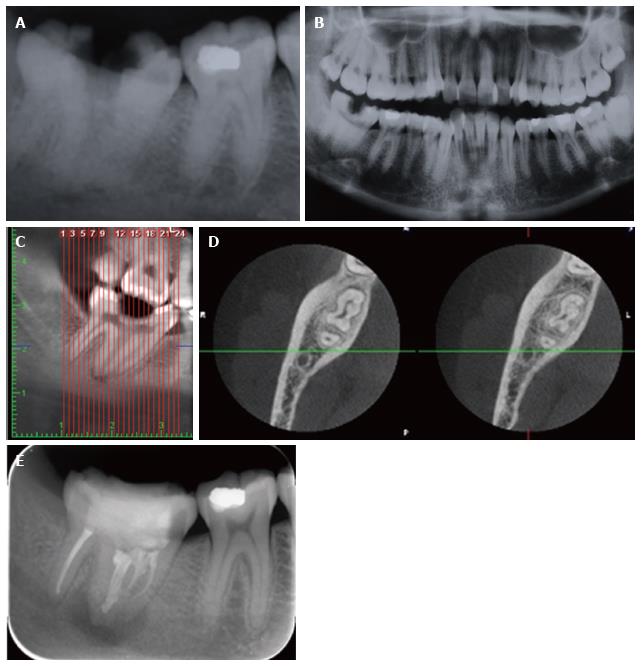
Figure 4 Radiological assessment of right mandibular 2nd molar tooth.
A: On periapical radiography, presence of caries and a periapical lesion was detected in a fused right mandibular 2nd molar tooth; B: The contrary left mandibular second molar was normal in shape on panoramic radiography; Panoramic CBCT (C) and axial CBCT (D) images showed that mandibular 2nd and 3rd molars were fused; E: The tooth was re-evaluated six months later with periapical radiography. The same periapical and CBCT units and settings with Case 1, 2, and 3 were used. CBCT: Cone beam computed tomography.
- Citation: Yılmaz F, Kamburoglu K, Yeta NY, Öztan MD. Cone beam computed tomography aided diagnosis and treatment of endodontic cases: Critical analysis. World J Radiol 2016; 8(7): 716-725
- URL: https://www.wjgnet.com/1949-8470/full/v8/i7/716.htm
- DOI: https://dx.doi.org/10.4329/wjr.v8.i7.716


