
















©The Author(s) 2016.
World J Radiol. Jul 28, 2016; 8(7): 700-706
Published online Jul 28, 2016. doi: 10.4329/wjr.v8.i7.700
Published online Jul 28, 2016. doi: 10.4329/wjr.v8.i7.700
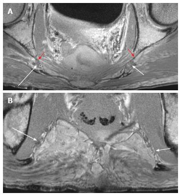
Figure 5 Male pelvic block-premarking.
Contiguous axial T1W images show the right (large arrows) and left (small arrows) pudendal nerves at the ischial spine (A) and Alcock’s canal (B). Corresponding coronal T1W image (C) shows suboptimal identification of the nerves (arrows) in the Alcock’s canal. Block arrows - sacrospinous ligaments.
- Citation: Chhabra A, McKenna CA, Wadhwa V, Thawait GK, Carrino JA, Lees GP, Dellon AL. 3T magnetic resonance neurography of pudendal nerve with cadaveric dissection correlation. World J Radiol 2016; 8(7): 700-706
- URL: https://www.wjgnet.com/1949-8470/full/v8/i7/700.htm
- DOI: https://dx.doi.org/10.4329/wjr.v8.i7.700