Copyright
©The Author(s) 2016.
World J Radiol. Jun 28, 2016; 8(6): 581-587
Published online Jun 28, 2016. doi: 10.4329/wjr.v8.i6.581
Published online Jun 28, 2016. doi: 10.4329/wjr.v8.i6.581
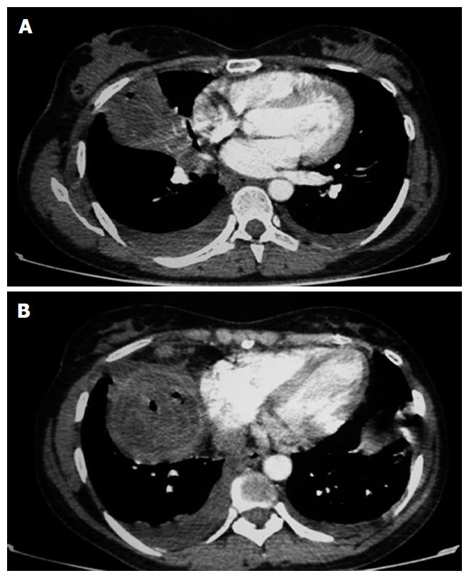
Figure 7 Complicated hydatid cyst showing ill defined wall with small air focus and consolidation in adjacent lung parenchyma (A) and posteroinferior to this cyst (B).
B showed air foci with serpingenous hyperattenuating membranes (“serpent sign”). Also note presence of mild bilateral pleural effusion, which was likely reactive.
- Citation: Garg MK, Sharma M, Gulati A, Gorsi U, Aggarwal AN, Agarwal R, Khandelwal N. Imaging in pulmonary hydatid cysts. World J Radiol 2016; 8(6): 581-587
- URL: https://www.wjgnet.com/1949-8470/full/v8/i6/581.htm
- DOI: https://dx.doi.org/10.4329/wjr.v8.i6.581


