Copyright
©The Author(s) 2016.
World J Radiol. Jun 28, 2016; 8(6): 571-580
Published online Jun 28, 2016. doi: 10.4329/wjr.v8.i6.571
Published online Jun 28, 2016. doi: 10.4329/wjr.v8.i6.571
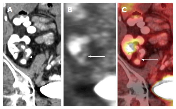
Figure 22 Coronal positron emission tomography computed tomography images of a patient with quiescent Crohn’s disease.
There is focal thickening of the distal ileum on the computed tomography component. This area of focal thickening does not show any fluorodeoxyglucose uptake on the positron emission tomography component, indicative that there is no significant associated regional inflammation and that this is likely a chronic stricture. Images used with permission from Dr. Martin O’Connell.
- Citation: Stanley E, Moriarty HK, Cronin CG. Advanced multimodality imaging of inflammatory bowel disease in 2015: An update. World J Radiol 2016; 8(6): 571-580
- URL: https://www.wjgnet.com/1949-8470/full/v8/i6/571.htm
- DOI: https://dx.doi.org/10.4329/wjr.v8.i6.571


