Copyright
©The Author(s) 2016.
World J Radiol. Jun 28, 2016; 8(6): 571-580
Published online Jun 28, 2016. doi: 10.4329/wjr.v8.i6.571
Published online Jun 28, 2016. doi: 10.4329/wjr.v8.i6.571
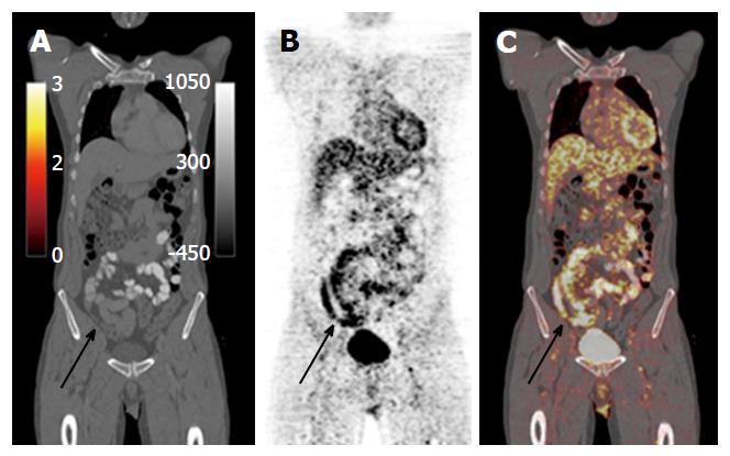
Figure 21 Oral contrast enhanced positron emission tomography computed tomography images in a patient with symptomatic Crohn’s disease shows nonspecific thickening of the distal and terminal ileum on the computed tomography component (A), with corresponding avid fluorodeoxyglucose uptake on the positron emission tomography component (B and C), indicative of acute inflammation.
- Citation: Stanley E, Moriarty HK, Cronin CG. Advanced multimodality imaging of inflammatory bowel disease in 2015: An update. World J Radiol 2016; 8(6): 571-580
- URL: https://www.wjgnet.com/1949-8470/full/v8/i6/571.htm
- DOI: https://dx.doi.org/10.4329/wjr.v8.i6.571


