Copyright
©The Author(s) 2016.
World J Radiol. Jun 28, 2016; 8(6): 571-580
Published online Jun 28, 2016. doi: 10.4329/wjr.v8.i6.571
Published online Jun 28, 2016. doi: 10.4329/wjr.v8.i6.571
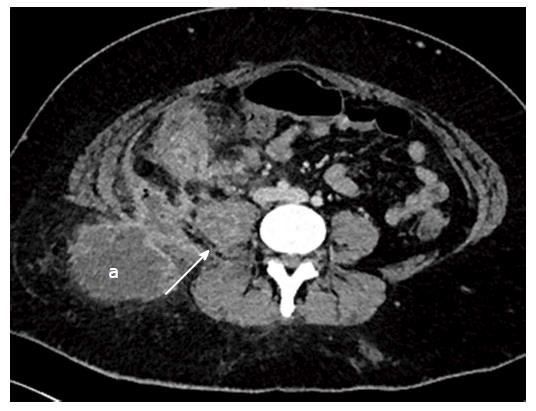
Figure 17 Axial contrast enhanced images of a 29-year-old lady with fistulating Crohn’s disease who presented with a mass in the right gluteal region, and raised CRP.
Computed tomography (CT) demonstrates ileocaecal inflammation associated with an abscess in right psoas (white arrow), extending to the posterior abdominal wall and the subcutaneous fat of the right gluteal region (a). Multiple pockets of air within the abscess indicate a communication between the bowel and the abscess. A pigtail drain was inserted the intra-abdominal component of the collection under CT guidance.
- Citation: Stanley E, Moriarty HK, Cronin CG. Advanced multimodality imaging of inflammatory bowel disease in 2015: An update. World J Radiol 2016; 8(6): 571-580
- URL: https://www.wjgnet.com/1949-8470/full/v8/i6/571.htm
- DOI: https://dx.doi.org/10.4329/wjr.v8.i6.571


