Copyright
©The Author(s) 2016.
World J Radiol. Jun 28, 2016; 8(6): 556-570
Published online Jun 28, 2016. doi: 10.4329/wjr.v8.i6.556
Published online Jun 28, 2016. doi: 10.4329/wjr.v8.i6.556
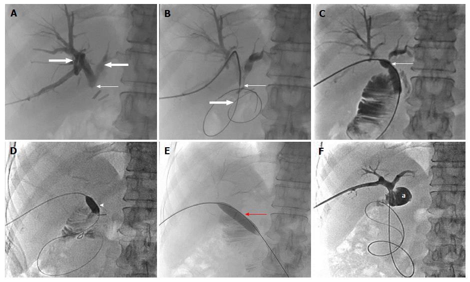
Figure 10 Fifty-four-year-old female, known case of extrahepatic portal vein obstruction with portal cavernoma cholangiopathy, status post hepatico-jejunostomy.
A: PTC showing complete cut-off at the HJ anastomotic site (thin arrow) with upstream bilobar biliary dilatation (block arrows); B: Fluoroscopic spot image showing the anastomotic site stricture (thin arrow) being negotiated using hydrophilic guide wire (block arrow); C: PTC with the help of conventional balloon showing resistent remant stricture at the anastomotic site in the form of persistent waist (thin arrow); D-F: Fluoroscopic spot images during PTC with combined use of cutting balloon (arrow head) and conventional balloon (red arrow) showing complete disappearance of stricture waist resulting into free flow of contrast into jejunal loop (a). PTC: Percutaneous transhepatic cholangiogram; HJ: Hepatico-jejunostomy.
- Citation: Pargewar SS, Desai SN, Rajesh S, Singh VP, Arora A, Mukund A. Imaging and radiological interventions in extra-hepatic portal vein obstruction. World J Radiol 2016; 8(6): 556-570
- URL: https://www.wjgnet.com/1949-8470/full/v8/i6/556.htm
- DOI: https://dx.doi.org/10.4329/wjr.v8.i6.556


