
















©The Author(s) 2016.
World J Radiol. May 28, 2016; 8(5): 484-500
Published online May 28, 2016. doi: 10.4329/wjr.v8.i5.484
Published online May 28, 2016. doi: 10.4329/wjr.v8.i5.484
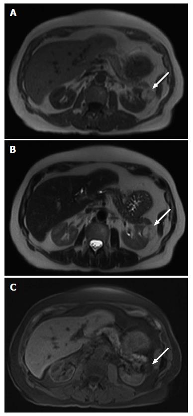
Figure 9 A 81-year-old male with a macroscopic fat containing renal angiomyolipoma.
A: On an axial T1-weighted magnetic resonance image. The ovoid lesion (arrow) in the left kidney shows uniform high T1 signal intensity; B: On an axial T2-weighted magnetic resonance image. The ovoid lesion (arrow) in the left kidney shows uniform high T2 signal intensity; C: On an axial fat suppressed T1-weighted magnetic resonance image. The ovoid lesion (arrow) in the left kidney which previously demonstrated uniform high T1 and T2 signal intensities now shows uniform signal loss.
- Citation: Low G, Huang G, Fu W, Moloo Z, Girgis S. Review of renal cell carcinoma and its common subtypes in radiology. World J Radiol 2016; 8(5): 484-500
- URL: https://www.wjgnet.com/1949-8470/full/v8/i5/484.htm
- DOI: https://dx.doi.org/10.4329/wjr.v8.i5.484