Copyright
©The Author(s) 2016.
World J Radiol. Mar 28, 2016; 8(3): 298-307
Published online Mar 28, 2016. doi: 10.4329/wjr.v8.i3.298
Published online Mar 28, 2016. doi: 10.4329/wjr.v8.i3.298
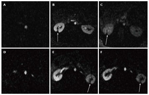
Figure 3 Selected 2D perfusion magnetic resonance images acquired from two representative animals.
The images show the arrival of Gd-DTPA bolus (A, D) in the aorta and 20-120 s (B, E and C, F) in the kidneys. Arrows denote the hypoperfused ablated lesions.
- Citation: Saeed M, Krug R, Do L, Hetts SW, Wilson MW. Renal ablation using magnetic resonance-guided high intensity focused ultrasound: Magnetic resonance imaging and histopathology assessment. World J Radiol 2016; 8(3): 298-307
- URL: https://www.wjgnet.com/1949-8470/full/v8/i3/298.htm
- DOI: https://dx.doi.org/10.4329/wjr.v8.i3.298


