
















©The Author(s) 2016.
World J Radiol. Mar 28, 2016; 8(3): 240-254
Published online Mar 28, 2016. doi: 10.4329/wjr.v8.i3.240
Published online Mar 28, 2016. doi: 10.4329/wjr.v8.i3.240
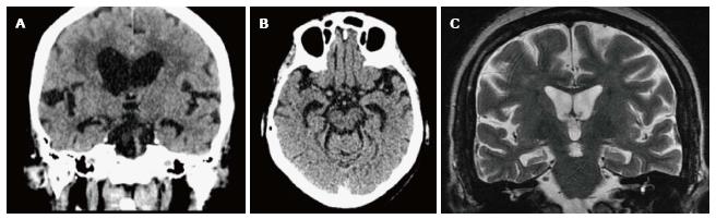
Figure 1 Hippocampal atrophy in an Alzheimer’s disease patient.
A: Computed tomography axial; B: Coronal images; C: Medial temporal lobe atrophy on magnetic resonance imaging (not the same patient).
- Citation: Narayanan L, Murray AD. What can imaging tell us about cognitive impairment and dementia? World J Radiol 2016; 8(3): 240-254
- URL: https://www.wjgnet.com/1949-8470/full/v8/i3/240.htm
- DOI: https://dx.doi.org/10.4329/wjr.v8.i3.240