Copyright
©The Author(s) 2016.
World J Radiol. Feb 28, 2016; 8(2): 159-173
Published online Feb 28, 2016. doi: 10.4329/wjr.v8.i2.159
Published online Feb 28, 2016. doi: 10.4329/wjr.v8.i2.159
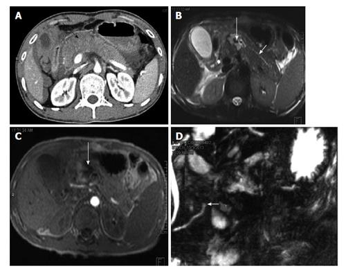
Figure 12 A 35-year-old man with history of road traffic accident.
CECT axial image (A) shows bulky hypoattenuating pancreas (black arrow) with peripancreatic fluid. No definite laceration was seen on CT and patient was kept on conservative management. MRI done 4 d later show hematoma/collection in neck of pancreas (long white arrows B and C). The duct was seen to communicate with the hematoma and MRCP showed cut off of duct at site of injury (short white arrow D). The patient subsequently underwent distal pancreatectomy. CECT: Contrast enhanced computed tomography; MRI: Magnetic resonance imaging; MRCP: Magnetic resonance pancreatography; CT: Computed tomography.
- Citation: Kumar A, Panda A, Gamanagatti S. Blunt pancreatic trauma: A persistent diagnostic conundrum? World J Radiol 2016; 8(2): 159-173
- URL: https://www.wjgnet.com/1949-8470/full/v8/i2/159.htm
- DOI: https://dx.doi.org/10.4329/wjr.v8.i2.159


