Copyright
©The Author(s) 2016.
World J Radiol. Feb 28, 2016; 8(2): 117-123
Published online Feb 28, 2016. doi: 10.4329/wjr.v8.i2.117
Published online Feb 28, 2016. doi: 10.4329/wjr.v8.i2.117
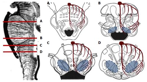
Figure 1 Schematic diagram of the brainstem vasculature.
A: Rostral pons; B: Caudal pons; C: Rostral medulla oblongata; D: Caudal medulla oblongata. Basilar artery, the terminal paramedian, short circumferential and long circumferential arteries are depicted. Blue shaded areas represent the tegmental watershed areas that are most frequently affected in neonates and infants with dorsal brainstem syndrome and a history of hypoxic-ischemic encephalopathy.
- Citation: Quattrocchi CC, Fariello G, Longo D. Brainstem tegmental lesions in neonates with hypoxic-ischemic encephalopathy: Magnetic resonance diagnosis and clinical outcome. World J Radiol 2016; 8(2): 117-123
- URL: https://www.wjgnet.com/1949-8470/full/v8/i2/117.htm
- DOI: https://dx.doi.org/10.4329/wjr.v8.i2.117


