
















©The Author(s) 2016.
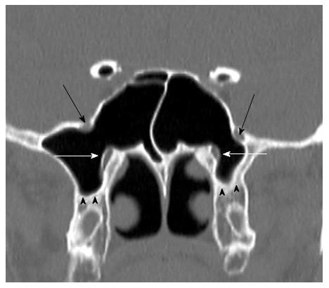
Figure 7 Coronal reformatted computerized tomography image shows bilaterally maxillary nerves (black arrows) and vidian nerves (white arows) protrusion into sphenoid sinus.
Additionally shows bilaterally pterygoid process pnematization (black arrow heads).
- Citation: Dasar U, Gokce E. Evaluation of variations in sinonasal region with computed tomography. World J Radiol 2016; 8(1): 98-108
- URL: https://www.wjgnet.com/1949-8470/full/v8/i1/98.htm
- DOI: https://dx.doi.org/10.4329/wjr.v8.i1.98