
















©The Author(s) 2016.
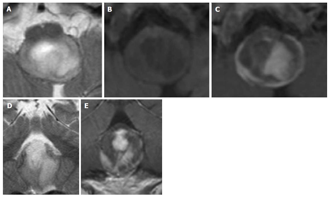
Figure 16 Pylocitic astrocytoma.
A 2-year-old boy with vomiting, respiratory distress and a dorsal exophytic medullary glioma Axial (A) and Coronal (D) T2 weighted images show a solid-cystic mass with an epicenter at the level of the medulla oblongata. Axial unenhanced (B) and axial (C) and coronal (E) enhanced T1 weighted images show inhomogeneous contrast enhancement.
- Citation: Quattrocchi CC, Errante Y, Rossi Espagnet MC, Galassi S, Della Sala SW, Bernardi B, Fariello G, Longo D. Magnetic resonance imaging differential diagnosis of brainstem lesions in children. World J Radiol 2016; 8(1): 1-20
- URL: https://www.wjgnet.com/1949-8470/full/v8/i1/1.htm
- DOI: https://dx.doi.org/10.4329/wjr.v8.i1.1