Copyright
©The Author(s) 2016.
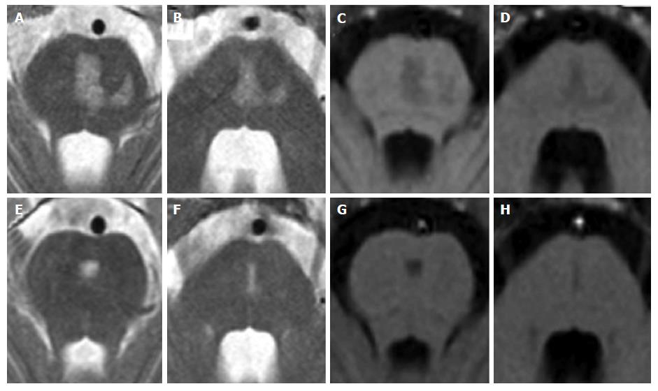
Figure 7 Central pontine myelinolysis.
A 11-year-old boy with lymphoma after a rapid correction of hyponatremia. Axial T2-weighted (A and B) and T1 weighted (C and D) images show signal alterations of the central pons. Note the incomplete trident shape and the sparing of the peripheral zones of the pons. Two years later, MR images (E to H) show minimal residual pontine alterations with absence of symptoms.
- Citation: Quattrocchi CC, Errante Y, Rossi Espagnet MC, Galassi S, Della Sala SW, Bernardi B, Fariello G, Longo D. Magnetic resonance imaging differential diagnosis of brainstem lesions in children. World J Radiol 2016; 8(1): 1-20
- URL: https://www.wjgnet.com/1949-8470/full/v8/i1/1.htm
- DOI: https://dx.doi.org/10.4329/wjr.v8.i1.1


