
















©The Author(s) 2015.
World J Radiol. Sep 28, 2015; 7(9): 253-265
Published online Sep 28, 2015. doi: 10.4329/wjr.v7.i9.253
Published online Sep 28, 2015. doi: 10.4329/wjr.v7.i9.253
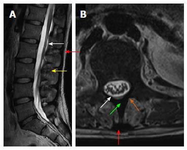
Figure 2 Magnetic resonance anatomy of the spine.
T2 weighted sagittal magnetic resonance (MR) image (A) and T2 weighted axial MR image (B) show posterior ligamentous complex with ligamentumflavum (white arrow), interspinous ligament (yellow arrow) supraspinous ligament (red arrow), spinous process (green arrow) and lamina (orange arrow).
- Citation: Gamanagatti S, Rathinam D, Rangarajan K, Kumar A, Farooque K, Sharma V. Imaging evaluation of traumatic thoracolumbar spine injuries: Radiological review. World J Radiol 2015; 7(9): 253-265
- URL: https://www.wjgnet.com/1949-8470/full/v7/i9/253.htm
- DOI: https://dx.doi.org/10.4329/wjr.v7.i9.253