Copyright
©The Author(s) 2015.
World J Radiol. Jun 28, 2015; 7(6): 110-127
Published online Jun 28, 2015. doi: 10.4329/wjr.v7.i6.110
Published online Jun 28, 2015. doi: 10.4329/wjr.v7.i6.110
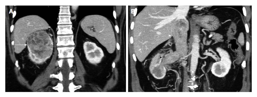
Figure 12 The 64-year-old man with clear cell renal cell carcinoma of the right kidney, invading the renal vein and the inferior vena cava (stage T3b, grade 3).
A: Coronal multiplanar reformations during the corticomedullary phase depicts large, inhomegeneously enhancing right renal tumor (arrow); B: Coronal 3D-display with maximum intensity projection technique during the same phase shows neoplastic thrombus invading left renal vein and the inferior vena cava (arrowheads). Coronal reformations clearly show venous invasion extending below the level of the diaphragm. Perinephric stranding and abnormal vessels are detected in the ipsilateral perinephric space, although pathology was negative for perinephric fat invasion.
- Citation: Tsili AC, Argyropoulou MI. Advances of multidetector computed tomography in the characterization and staging of renal cell carcinoma. World J Radiol 2015; 7(6): 110-127
- URL: https://www.wjgnet.com/1949-8470/full/v7/i6/110.htm
- DOI: https://dx.doi.org/10.4329/wjr.v7.i6.110


