Copyright
©The Author(s) 2015.
World J Radiol. Jun 28, 2015; 7(6): 110-127
Published online Jun 28, 2015. doi: 10.4329/wjr.v7.i6.110
Published online Jun 28, 2015. doi: 10.4329/wjr.v7.i6.110
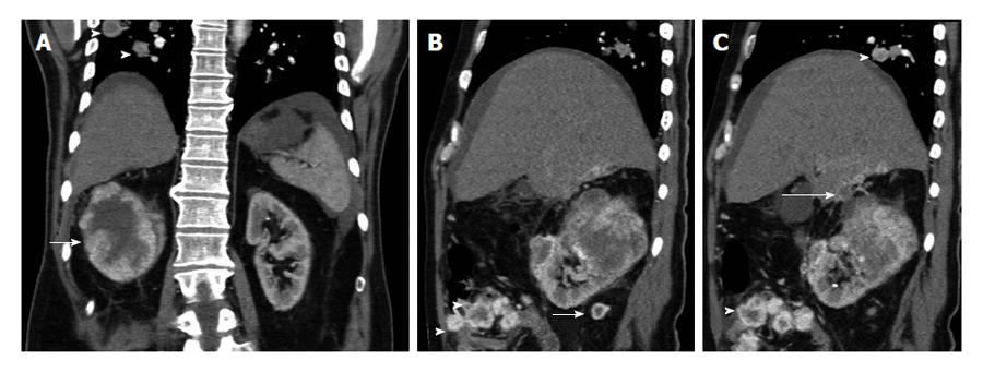
Figure 3 The 52-year-old man with advanced-stage clear cell renal cell carcinoma of the right kidney.
Contrast-enhanced (A) coronal and (B and C) sagittal reformations during the corticomedullar phase show a large right renal tumor (arrow), strongly and inhomogeneously enhancing. Central hypodense parts within malignancy corresponded to areas of necrosis on histology. There is perinephric stranding and contrast-enhancing nodules in the perinephric fat (long arrow, B), a finding strongly suggestive for perinephric fat invasion. The tumor is seen extending and invading the undersurface of the liver (long arrow, C). Lung metastases are detected (arrowheads, A and C). There is also a small amount of ascites and nodular peritoneal masses (arrowheads, B), with heterogeneous enhancement, identical to that of the primary neoplasm, findings suggestive of peritoneal metastases. Peritoneal metastases from renal cell carcinoma (RCC) are extremely rare. Neoplastic invasion of the peritoneum by RCC may occur either, by contiguous spread of renal tumor through the renal capsule, the anterior renal fascia and the posterior parietal peritoneum, or via tumoral emboli.
- Citation: Tsili AC, Argyropoulou MI. Advances of multidetector computed tomography in the characterization and staging of renal cell carcinoma. World J Radiol 2015; 7(6): 110-127
- URL: https://www.wjgnet.com/1949-8470/full/v7/i6/110.htm
- DOI: https://dx.doi.org/10.4329/wjr.v7.i6.110


