Copyright
©The Author(s) 2015.
World J Radiol. Jun 28, 2015; 7(6): 110-127
Published online Jun 28, 2015. doi: 10.4329/wjr.v7.i6.110
Published online Jun 28, 2015. doi: 10.4329/wjr.v7.i6.110
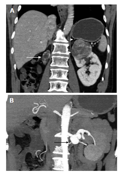
Figure 2 The 70-year-old man with clear cell renal cell carcinoma of the left solitary functioning kidney (stage T1b, grade 2).
A: Post-contrast enhanced coronal multiplanar reformation during the corticomedullary phase depicts left upper pole renal mass, strongly and heterogeneously enhancing, after contrast material administration. A thin hyperdense rim (arrowheads) is detected around the tumor, proved to correspond to fibrous pseudocapsule on pathology. Atrophic right kidney (arrow); B: Coronal 3D-reconstruction during the same phase, using maximum intensity projection algorithm shows left renal artery aneurysm (arrow).
- Citation: Tsili AC, Argyropoulou MI. Advances of multidetector computed tomography in the characterization and staging of renal cell carcinoma. World J Radiol 2015; 7(6): 110-127
- URL: https://www.wjgnet.com/1949-8470/full/v7/i6/110.htm
- DOI: https://dx.doi.org/10.4329/wjr.v7.i6.110


