Copyright
©The Author(s) 2015.
World J Radiol. May 28, 2015; 7(5): 104-109
Published online May 28, 2015. doi: 10.4329/wjr.v7.i5.104
Published online May 28, 2015. doi: 10.4329/wjr.v7.i5.104
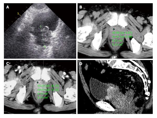
Figure 4 A 72-year-old male patient presented with one month of dysuria.
A: Prostate enlarged to 4.0 cm × 5.1 cm × 4.2 cm with incomplete envelope, irregular shape and heterogeneous echo texture; B: A pre-enhanced computed tomography (CT) scan image of the pelvis showed a marked enlarged prostatic tumor invading to bladder, seminal vesicle; C: The enlarged prostate was inhomogeneous moderately enhancement in arterial phase; D: Coronal oblique multiplanar reconstruction of enhanced CT scan shows the enhanced inhomogeneous prostate with high attenuation protruding into bladder and unclear with surrounding tissue.
- Citation: He HQ, Fan SF, Xu Q, Chen ZJ, Li Z. Diagnosis of prostatic neuroendocrine carcinoma: Two cases report and literature review. World J Radiol 2015; 7(5): 104-109
- URL: https://www.wjgnet.com/1949-8470/full/v7/i5/104.htm
- DOI: https://dx.doi.org/10.4329/wjr.v7.i5.104


