Copyright
©The Author(s) 2015.
World J Radiol. May 28, 2015; 7(5): 100-103
Published online May 28, 2015. doi: 10.4329/wjr.v7.i5.100
Published online May 28, 2015. doi: 10.4329/wjr.v7.i5.100
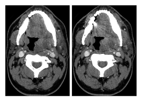
Figure 1 Axial computed tomography images at the level of the oropharynx prior to incision, drainage and silver nitrate application demonstrate swelling and enlargement of the left palatine tonsil and peritonsillar tissues with heterogeneous relatively low attenuation areas.
- Citation: Livingstone D, Alghonaim Y, Jowett N, Sela E, Mlynarek A, Forghani R. Silver nitrate mimicking a foreign body in the pharyngeal mucosal space. World J Radiol 2015; 7(5): 100-103
- URL: https://www.wjgnet.com/1949-8470/full/v7/i5/100.htm
- DOI: https://dx.doi.org/10.4329/wjr.v7.i5.100


