
















©The Author(s) 2015.
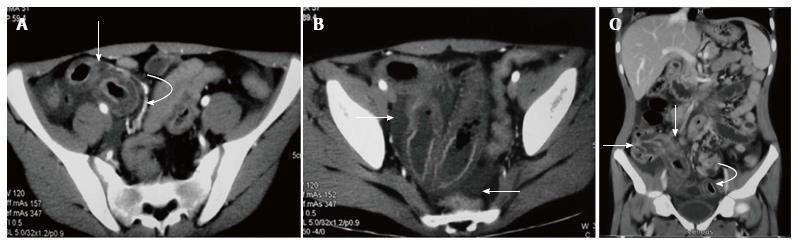
Figure 1 Bowel involvement in Henoch-Schonlein purpura in a 15-year-old male.
A: Contrast enhanced axial computed tomography (CT) image in the arterial phase shows circumferential wall thickening involving a distal ileal loop (arrow) with mural stratification. Engorged mesenteric vessels are seen adjacent to the thickened bowel (curved arrow); B: Axial CT image at the level of pelvis shows free fluid (arrows) and thickening of ileal loops; C: Coronal multiplanar reformatted image shows long segment thickening involving terminal ileum and ascending colon (straight arrows). Another segment of distal ileal loop also shows wall thickening (curved arrow).
- Citation: Prathiba Rajalakshmi P, Srinivasan K. Gastrointestinal manifestations of Henoch-Schonlein purpura: A report of two cases. World J Radiol 2015; 7(3): 66-69
- URL: https://www.wjgnet.com/1949-8470/full/v7/i3/66.htm
- DOI: https://dx.doi.org/10.4329/wjr.v7.i3.66