
















©The Author(s) 2015.
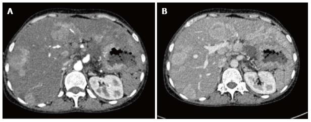
Figure 3 Arterial (A) and venous (B) phases of multiphase contrast enhanced axial computed tomography images showing multiple arterial enhancing focal lesions in liver with no significant wash out in venous phase.
- Citation: Prasad T, Madhusudhan K, Srivastava DN, Dash NR, Gupta AK. Transarterial chemoembolization for liver metastases from solid pseudopapillary epithelial neoplasm of pancreas: A case report. World J Radiol 2015; 7(3): 61-65
- URL: https://www.wjgnet.com/1949-8470/full/v7/i3/61.htm
- DOI: https://dx.doi.org/10.4329/wjr.v7.i3.61