Copyright
©The Author(s) 2015.
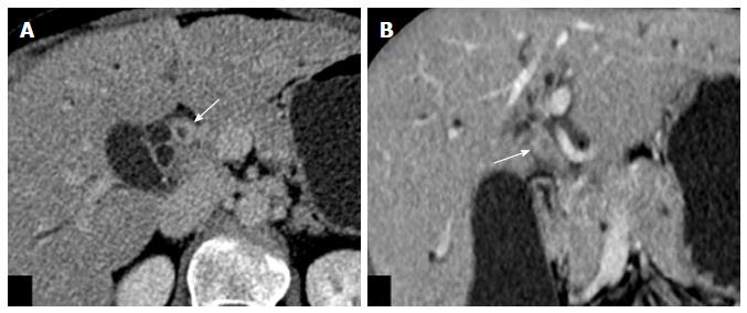
Figure 9 Axial (A) and coronal (B) images of venous phase of computed tomography scan of periductal infiltrating type of hilar cholangiocarcinoma showing wall thickening and enhancement of proximal common hepatic duct (arrow) causing luminal obstruction.
- Citation: Madhusudhan KS, Gamanagatti S, Gupta AK. Imaging and interventions in hilar cholangiocarcinoma: A review. World J Radiol 2015; 7(2): 28-44
- URL: https://www.wjgnet.com/1949-8470/full/v7/i2/28.htm
- DOI: https://dx.doi.org/10.4329/wjr.v7.i2.28


