
















©The Author(s) 2015.
World J Radiol. Dec 28, 2015; 7(12): 459-474
Published online Dec 28, 2015. doi: 10.4329/wjr.v7.i12.459
Published online Dec 28, 2015. doi: 10.4329/wjr.v7.i12.459
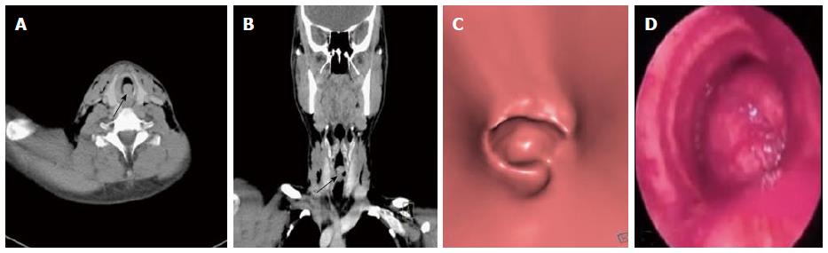
Figure 6 Adenoid cystic carcinoma of trachea.
Axial (A) and coronal MPR (B) images show polypoidal mass with smooth outline within the trachea in subglottic region causing near complete attenuation of the airway (arrow). Virtual bronchoscopy (C) shows intraluminal smooth mass within trachea similar to that seen on conventional bronchoscopy (D). MPR: Multiplanar reformation.
- Citation: Jugpal TS, Garg A, Sethi GR, Daga MK, Kumar J. Multi-detector computed tomography imaging of large airway pathology: A pictorial review. World J Radiol 2015; 7(12): 459-474
- URL: https://www.wjgnet.com/1949-8470/full/v7/i12/459.htm
- DOI: https://dx.doi.org/10.4329/wjr.v7.i12.459