
















©The Author(s) 2015.
World J Radiol. Dec 28, 2015; 7(12): 448-458
Published online Dec 28, 2015. doi: 10.4329/wjr.v7.i12.448
Published online Dec 28, 2015. doi: 10.4329/wjr.v7.i12.448
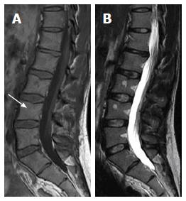
Figure 9 Sagittal T1W (A) and T2W (B) images of 33-year-old male showing focal geographic low signal intensity patches targeting LV3 and LV4 bodies centers as well as around basi-vertebral veins.
These patches still of high-signal intensity on T2W image. Note, the central fat spot (white arrow) and fuzzy margins of LV4 lesion inferring benignity features are consistent with focal nodular marrow reconversion.
- Citation: Nouh MR, Eid AF. Magnetic resonance imaging of the spinal marrow: Basic understanding of the normal marrow pattern and its variant. World J Radiol 2015; 7(12): 448-458
- URL: https://www.wjgnet.com/1949-8470/full/v7/i12/448.htm
- DOI: https://dx.doi.org/10.4329/wjr.v7.i12.448