Copyright
©The Author(s) 2015.
World J Radiol. Dec 28, 2015; 7(12): 438-447
Published online Dec 28, 2015. doi: 10.4329/wjr.v7.i12.438
Published online Dec 28, 2015. doi: 10.4329/wjr.v7.i12.438
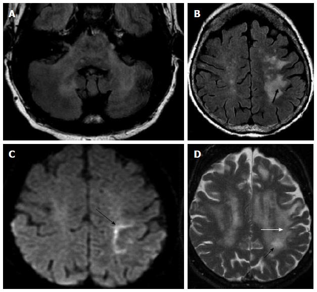
Figure 3 Progressive multifocal leucoencephalopathy.
HIV patient with + JC virus on PCR-CSF. Bilateral involvement of MCP on axial FLAIR (A); with no enhancement (not shown); Axial FLAIR (B) and axial DWI (C) images show asymmetric confluent areas of increased signal in the subcortical WM with involvement of the u-fibers (arrow) and linear “edge” on DWI (arrow on C) consistent with advancing demyelinated edge; D: Three months f/u images better depict involvement of u-fibers (white arrow) as well as progression of WM disease with formation of small central cavitation/micro cyst, characteristic of PML (black arrow). PML: Progressive multifocal leucoencephalopathy; MCP: Middle cerebellar peduncles; CSF: Cerebrospinal fluid; DWI: Diffusion weighted-imaging; PCR: Polymerase chain reaction; HIV: Human immunodeficiency virus.
- Citation: Morales H, Tomsick T. Middle cerebellar peduncles: Magnetic resonance imaging and pathophysiologic correlate. World J Radiol 2015; 7(12): 438-447
- URL: https://www.wjgnet.com/1949-8470/full/v7/i12/438.htm
- DOI: https://dx.doi.org/10.4329/wjr.v7.i12.438


