Copyright
©The Author(s) 2015.
World J Radiol. Nov 28, 2015; 7(11): 394-404
Published online Nov 28, 2015. doi: 10.4329/wjr.v7.i11.394
Published online Nov 28, 2015. doi: 10.4329/wjr.v7.i11.394
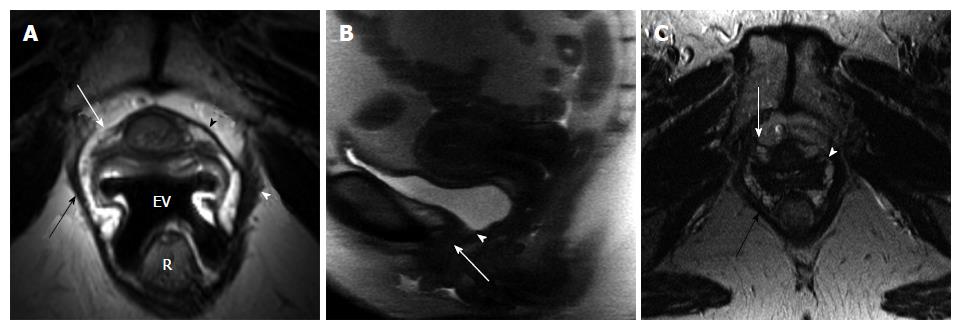
Figure 2 51-year-old woman post two uneventful vaginal deliveries, body mass index: 24.
3. A: Axial oblique T2-weighted image at the mid urethra level (at 50% length from the internal meatus, about 1.2 cm), perpendicular to the axis of urethra, obtained with endovaginal placement of MRInnervu coil (EV) (TR/TE 3200/93 ms) shows disrupted attenuated right periurethral ligament (white arrow). Note intact left side of the periurethral ligament (black arrowhead) and its attachment to the puborectalis muscle (white arrowhead). Minimal asymmetric thinning of the right puborectalis muscle (black arrow); B: Sagittal SSFSE image (TR/TE 15000/78 ms) during strain shows hypermobility of the urethra (arrow). Note closure of bladder neck (arrowhead) during the urethral descent; C: Axial T2-weighted image of the pelvis at the level of mid urethra (at 50% from the internal meatus) obtained with pelvic coil (TR/TE 4666/85 ms) shows much less detail of the periurethral ligament compared to endovaginal MRI in A. It is difficult to appreciate the status of the ligament itself (white arrow) or its attachment (white arrowhead). Puborectalis muscle (black arrow) is well visualized. R: Rectum.
- Citation: Macura KJ, Thompson RE, Bluemke DA, Genadry R. Magnetic resonance imaging in assessment of stress urinary incontinence in women: Parameters differentiating urethral hypermobility and intrinsic sphincter deficiency. World J Radiol 2015; 7(11): 394-404
- URL: https://www.wjgnet.com/1949-8470/full/v7/i11/394.htm
- DOI: https://dx.doi.org/10.4329/wjr.v7.i11.394


