















©The Author(s) 2015.
World J Radiol. Oct 28, 2015; 7(10): 357-360
Published online Oct 28, 2015. doi: 10.4329/wjr.v7.i10.357
Published online Oct 28, 2015. doi: 10.4329/wjr.v7.i10.357
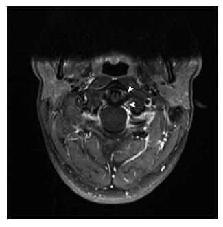
Figure 3 Follow-up magnetic resonance imaging.
High resolution contrast-enhanced 3T magnetic resonance imaging, fat-saturated gradient echo sequence. After physical therapy stabilization. Note the clear contrast enhancement in the periligamentous venous plexus (arrow head) and the symmetric space evaluation (white arrow).
- Citation: Kaufmann RA, Marzi I, Vogl TJ. Delayed diagnosis of isolated alar ligament rupture: A case report. World J Radiol 2015; 7(10): 357-360
- URL: https://www.wjgnet.com/1949-8470/full/v7/i10/357.htm
- DOI: https://dx.doi.org/10.4329/wjr.v7.i10.357