
















©The Author(s) 2015.
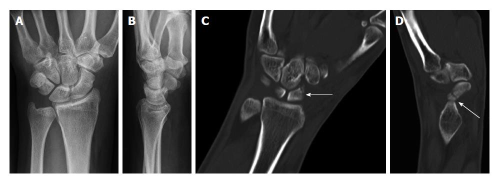
Figure 1 Non-dislocated fissural fracture of the scaphoid that had not been detected by initial conventional radiography (A and B).
Cross-sectional multidetector computed tomography clearly depicted the oblique fissural fracture line in the middle third of the scaphoid with coronal (C) and sagittal (D) reformation on the same day.
- Citation: Behzadi C, Karul M, Henes FO, Laqmani A, Catala-Lehnen P, Lehmann W, Nagel HD, Adam G, Regier M. Comparison of conventional radiography and MDCT in suspected scaphoid fractures. World J Radiol 2015; 7(1): 22-27
- URL: https://www.wjgnet.com/1949-8470/full/v7/i1/22.htm
- DOI: https://dx.doi.org/10.4329/wjr.v7.i1.22