
















©2014 Baishideng Publishing Group Inc.
World J Radiol. Sep 28, 2014; 6(9): 716-725
Published online Sep 28, 2014. doi: 10.4329/wjr.v6.i9.716
Published online Sep 28, 2014. doi: 10.4329/wjr.v6.i9.716
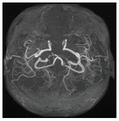
Figure 11 Time of Flight magnetic resonance angiography in a patient with tubercular meningitis.
Bilateral anterior cerebral arteries are not seen whereas bilateral middle cerebral arteries are markedly attenuated suggesting vasculitis. The patient presented with extensive cerebral infarction.
- Citation: Rangarajan K, Das CJ, Kumar A, Gupta AK. MRI in central nervous system infections: A simplified patterned approach. World J Radiol 2014; 6(9): 716-725
- URL: https://www.wjgnet.com/1949-8470/full/v6/i9/716.htm
- DOI: https://dx.doi.org/10.4329/wjr.v6.i9.716