Copyright
©2014 Baishideng Publishing Group Inc.
World J Radiol. Sep 28, 2014; 6(9): 716-725
Published online Sep 28, 2014. doi: 10.4329/wjr.v6.i9.716
Published online Sep 28, 2014. doi: 10.4329/wjr.v6.i9.716
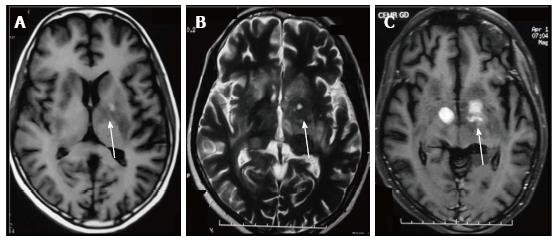
Figure 3 T1 (A), T2 (B) and Post gadolinium T1 (C) weighted images in an human immunodeficiency virus-positive patient with space occupying lesions in bilateral basal ganglia.
Differentials for this appearance in such a patient would include Toxoplasmosis, Cryptococcosis as well as central nervous system (CNS) lymphoma. This patient had CNS lymphoma.
- Citation: Rangarajan K, Das CJ, Kumar A, Gupta AK. MRI in central nervous system infections: A simplified patterned approach. World J Radiol 2014; 6(9): 716-725
- URL: https://www.wjgnet.com/1949-8470/full/v6/i9/716.htm
- DOI: https://dx.doi.org/10.4329/wjr.v6.i9.716


