
















©2014 Baishideng Publishing Group Inc.
World J Radiol. Sep 28, 2014; 6(9): 677-692
Published online Sep 28, 2014. doi: 10.4329/wjr.v6.i9.677
Published online Sep 28, 2014. doi: 10.4329/wjr.v6.i9.677
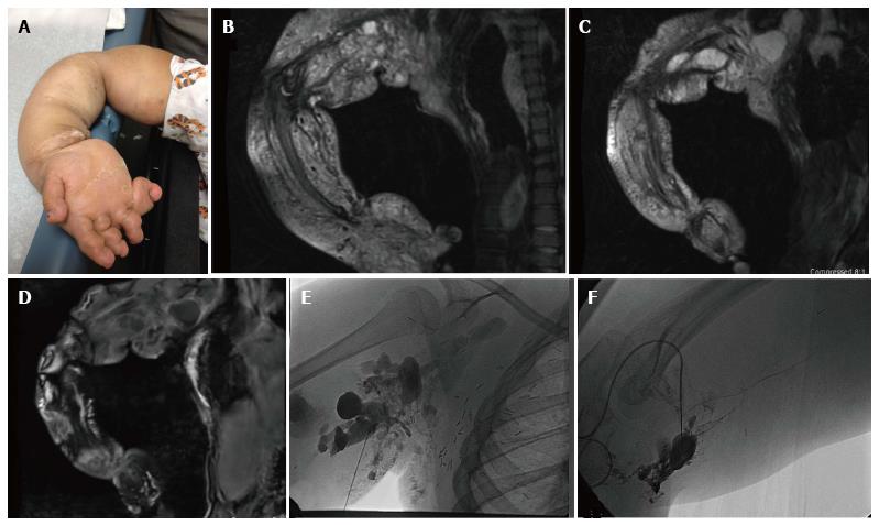
Figure 6 Ten-year-old girl with CLOVES syndrome.
Photograph (A) demonstrates right upper extremity overgrowth. Pre-treatment coronal MR images of the right upper extremity (B, C, D) demonstrate a large, combined macro/microcystic lymphatic malformation with venous lakes in the axilla and evidence of lipomatous overgrowth. Angiographic images (E, F) demonstrate direct puncture of the malformation followed by sclerosis with doxycycline.
- Citation: Nosher JL, Murillo PG, Liszewski M, Gendel V, Gribbin CE. Vascular anomalies: A pictorial review of nomenclature, diagnosis and treatment. World J Radiol 2014; 6(9): 677-692
- URL: https://www.wjgnet.com/1949-8470/full/v6/i9/677.htm
- DOI: https://dx.doi.org/10.4329/wjr.v6.i9.677