
















©2014 Baishideng Publishing Group Inc.
World J Radiol. Sep 28, 2014; 6(9): 657-668
Published online Sep 28, 2014. doi: 10.4329/wjr.v6.i9.657
Published online Sep 28, 2014. doi: 10.4329/wjr.v6.i9.657
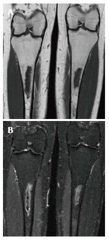
Figure 7 Medullary infarction.
Coronal T1 WI (A) image shows irregularly bordered areas of low SI within the medullary cavity of both tibiae. The same areas show peripheral serpigenous high SI on the coronal short tau inversion recovery (STIR) (B) image. The appearance is typical of an infarct.
- Citation: Simpson WL, Hermann G, Balwani M. Imaging of Gaucher disease. World J Radiol 2014; 6(9): 657-668
- URL: https://www.wjgnet.com/1949-8470/full/v6/i9/657.htm
- DOI: https://dx.doi.org/10.4329/wjr.v6.i9.657