Copyright
©2014 Baishideng Publishing Group Inc.
World J Radiol. Sep 28, 2014; 6(9): 657-668
Published online Sep 28, 2014. doi: 10.4329/wjr.v6.i9.657
Published online Sep 28, 2014. doi: 10.4329/wjr.v6.i9.657
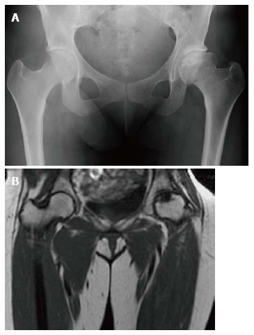
Figure 6 Osteonecrosis.
Frontal radiograph of the pelvis (A) shows avascular necrosis of the left femoral head. The femoral head has a flattened contour with sclerosis in the subcapsular areas. Note that the joint space is maintained. Coronal T1 WI (B) in the same patient again demonstrated an abnormal shape of the left femoral head with flattening superiorly. In the same area there is a focus of low SI indicating the devasularized bone.
- Citation: Simpson WL, Hermann G, Balwani M. Imaging of Gaucher disease. World J Radiol 2014; 6(9): 657-668
- URL: https://www.wjgnet.com/1949-8470/full/v6/i9/657.htm
- DOI: https://dx.doi.org/10.4329/wjr.v6.i9.657


