Copyright
©2014 Baishideng Publishing Group Inc.
World J Radiol. Aug 28, 2014; 6(8): 619-624
Published online Aug 28, 2014. doi: 10.4329/wjr.v6.i8.619
Published online Aug 28, 2014. doi: 10.4329/wjr.v6.i8.619
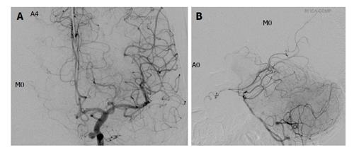
Figure 3 A 50-year-old patient with left skull base meningioma and an incidental left internal carotid artery aneurysm (Grade A4, M0; circle of Willis Type 1; balloon test occlusion negative).
No cross flow is visualized in the right middle cerebral artery by either left internal carotid artery (ICA) (A) or vertebral artery (VA) (B) injection. The right anterior cerebral artery is well visualized by left ICA injection (A) but not by VA injection (B). This patient complained of right facial paralysis and aphasia 8 min after the balloon occlusion was inflated.
- Citation: Kikuchi K, Yoshiura T, Hiwatashi A, Togao O, Yamashita K, Honda H. Balloon test occlusion of internal carotid artery: Angiographic findings predictive of results. World J Radiol 2014; 6(8): 619-624
- URL: https://www.wjgnet.com/1949-8470/full/v6/i8/619.htm
- DOI: https://dx.doi.org/10.4329/wjr.v6.i8.619


