Copyright
©2014 Baishideng Publishing Group Inc.
World J Radiol. Aug 28, 2014; 6(8): 619-624
Published online Aug 28, 2014. doi: 10.4329/wjr.v6.i8.619
Published online Aug 28, 2014. doi: 10.4329/wjr.v6.i8.619
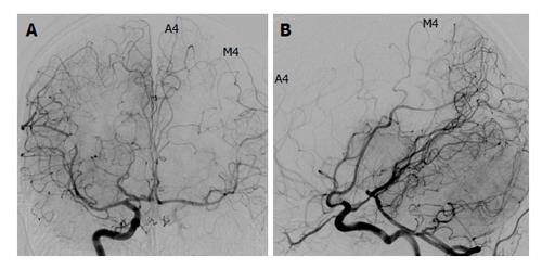
Figure 2 A 30-year-old patient with left skull base meningioma (Grade A4, M4; circle of Willis Type 3; balloon test occlusion negative).
In the right internal carotid artery injection, both the anterior cerebral artery and the middle cerebral artery on the left are well visualized by a cross flow via the anterior communicating artery (A) and the posterior communicating artery (B).
- Citation: Kikuchi K, Yoshiura T, Hiwatashi A, Togao O, Yamashita K, Honda H. Balloon test occlusion of internal carotid artery: Angiographic findings predictive of results. World J Radiol 2014; 6(8): 619-624
- URL: https://www.wjgnet.com/1949-8470/full/v6/i8/619.htm
- DOI: https://dx.doi.org/10.4329/wjr.v6.i8.619


