
















©2014 Baishideng Publishing Group Inc.
World J Radiol. Aug 28, 2014; 6(8): 530-537
Published online Aug 28, 2014. doi: 10.4329/wjr.v6.i8.530
Published online Aug 28, 2014. doi: 10.4329/wjr.v6.i8.530
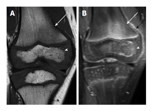
Figure 6 Early acute osteomyelitis.
A: Coronal T1-weighted image of the right knee of a 4-year-old male shows ill-defined areas of low T1 signal in the bone marrow in the lateral femoral metaphysis (arrow) and lateral epiphysis (arrowhead); B: Coronal T1-weighted post-contrast image in the same patient shows associated enhancement in these areas (white arrow and arrowhead) as well as some periosteal reaction, indicated by periosteal enhancement (black arrow).
- Citation: Pugmire BS, Shailam R, Gee MS. Role of MRI in the diagnosis and treatment of osteomyelitis in pediatric patients. World J Radiol 2014; 6(8): 530-537
- URL: https://www.wjgnet.com/1949-8470/full/v6/i8/530.htm
- DOI: https://dx.doi.org/10.4329/wjr.v6.i8.530