Copyright
©2014 Baishideng Publishing Group Inc.
World J Radiol. Jul 28, 2014; 6(7): 424-436
Published online Jul 28, 2014. doi: 10.4329/wjr.v6.i7.424
Published online Jul 28, 2014. doi: 10.4329/wjr.v6.i7.424
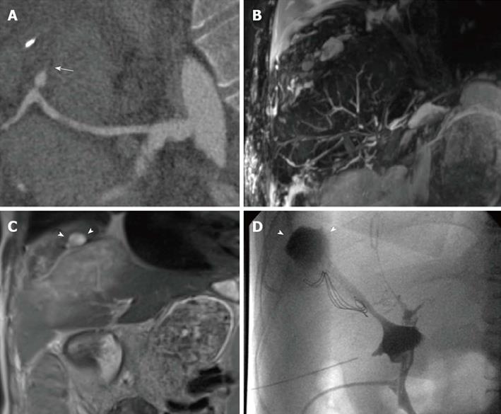
Figure 6 Multiple findings in a 28-year-old female patient transplanted for primary sclerosing cholangitis.
Because of the hepatic artery thrombosis shown on curved-reformatted CT image (arrow in A), the biliary tree appears as fragmented and anatomically ill-defined on a panoramic maximum intensity projection view (B). Coronal T2-weighted HASTE image (C) shows extensive, hyperintense ischemic damage of liver parenchyma, together with intrahepatic fluid collections (arrowheads) confirmed to be the effect of bile leakage on T-tube cholangiography (arrowheads in D).
- Citation: Girometti R, Cereser L, Bazzocchi M, Zuiani C. Magnetic resonance cholangiography in the assessment and management of biliary complications after OLT. World J Radiol 2014; 6(7): 424-436
- URL: https://www.wjgnet.com/1949-8470/full/v6/i7/424.htm
- DOI: https://dx.doi.org/10.4329/wjr.v6.i7.424


