Copyright
©2014 Baishideng Publishing Group Inc.
World J Radiol. Jul 28, 2014; 6(7): 424-436
Published online Jul 28, 2014. doi: 10.4329/wjr.v6.i7.424
Published online Jul 28, 2014. doi: 10.4329/wjr.v6.i7.424
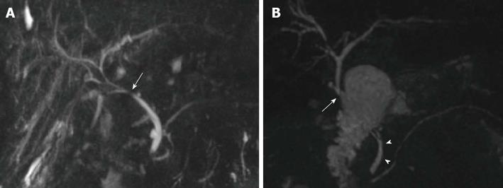
Figure 1 Biliary reconstructions variants after orthotopic liver transplantation illustrated by coronal maximum intensity projection reconstruction from 3D magnetic resonance cholangiography.
A: Choledocho-choledochostomy with mild donor-to-recipient discrepancy in ductal calibers giving prominence to the anastomotic site (arrow); B: Bilioenteric anastomosis (arrow) between donor’s common bile duct and a jejuneal loop. Note the recipient common bile duct remnant (arrowheads).
- Citation: Girometti R, Cereser L, Bazzocchi M, Zuiani C. Magnetic resonance cholangiography in the assessment and management of biliary complications after OLT. World J Radiol 2014; 6(7): 424-436
- URL: https://www.wjgnet.com/1949-8470/full/v6/i7/424.htm
- DOI: https://dx.doi.org/10.4329/wjr.v6.i7.424


