Copyright
©2014 Baishideng Publishing Group Inc.
World J Radiol. Jun 28, 2014; 6(6): 388-391
Published online Jun 28, 2014. doi: 10.4329/wjr.v6.i6.388
Published online Jun 28, 2014. doi: 10.4329/wjr.v6.i6.388
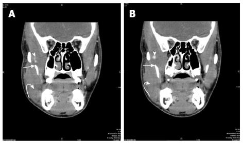
Figure 2 Three millimeter reconstructed contrast enhanced coronal computed tomography images of the face with soft tissue settings demonstrate swelling of the entire right temporalis muscle more prominent at its myotendinous insertion to the mandible (straight arrow).
There is also swelling of right masseter muscle (curved arrow). Small high density material is again seen in region of myotendinous insertion of right temporalis muscle suggesting hemorrhage (arrowhead).
- Citation: Naffaa LN, Tandon YK, Rubin M. Myotendinous rupture of temporalis muscle: A rare injury following seizure. World J Radiol 2014; 6(6): 388-391
- URL: https://www.wjgnet.com/1949-8470/full/v6/i6/388.htm
- DOI: https://dx.doi.org/10.4329/wjr.v6.i6.388


