Copyright
©2014 Baishideng Publishing Group Inc.
World J Radiol. Jun 28, 2014; 6(6): 374-380
Published online Jun 28, 2014. doi: 10.4329/wjr.v6.i6.374
Published online Jun 28, 2014. doi: 10.4329/wjr.v6.i6.374
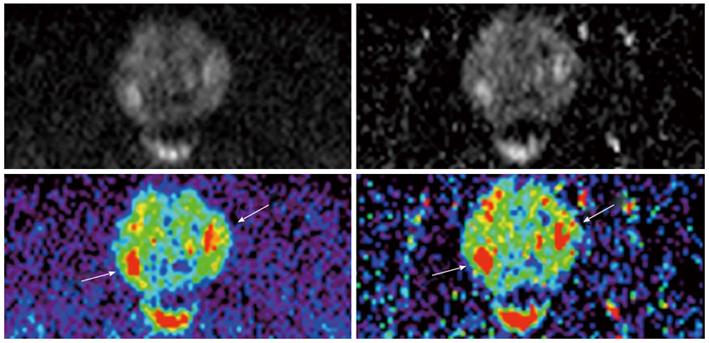
Figure 3 M-b1400 (left column) and C-b1400 (right column) images of the same patient.
Demarcation of two oval hyperintense suspicious lesions is very good on both images (arrows).
-
Citation: Bittencourt LK, Attenberger UI, Lima D, Strecker R, Oliveira A, Schoenberg SO, Gasparetto EL, Hausmann D. Feasibility study of computed
vs measured high b-value (1400 s/mm²) diffusion-weighted MR images of the prostate. World J Radiol 2014; 6(6): 374-380 - URL: https://www.wjgnet.com/1949-8470/full/v6/i6/374.htm
- DOI: https://dx.doi.org/10.4329/wjr.v6.i6.374


