Copyright
©2014 Baishideng Publishing Group Inc.
World J Radiol. Jun 28, 2014; 6(6): 355-365
Published online Jun 28, 2014. doi: 10.4329/wjr.v6.i6.355
Published online Jun 28, 2014. doi: 10.4329/wjr.v6.i6.355
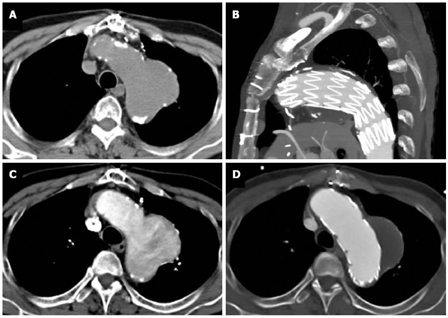
Figure 11 A 68-year-old with chest pain and long standing hypertension.
A saccular aneurysm is seen arising from the lateral aortic arch just distal to the origin of the left subclavian artery on this computed tomography aortogram. Atherosclerotic calcifications are seen at the periphery of the aneurysm (A) and there is heterogeneous contrast opacification secondary to turbulent flow on the arterial phase (B). No evidence of rupture is apparent. The aneurysm was excluded using an aortic stent graft as seen in the sagittal plane (C). The excluded sac has completely thrombosed with no endoleak as seen on an axial image (D).
- Citation: Hallinan JTPD, Anil G. Multi-detector computed tomography in the diagnosis and management of acute aortic syndromes. World J Radiol 2014; 6(6): 355-365
- URL: https://www.wjgnet.com/1949-8470/full/v6/i6/355.htm
- DOI: https://dx.doi.org/10.4329/wjr.v6.i6.355


