Copyright
©2014 Baishideng Publishing Group Inc.
World J Radiol. Jun 28, 2014; 6(6): 355-365
Published online Jun 28, 2014. doi: 10.4329/wjr.v6.i6.355
Published online Jun 28, 2014. doi: 10.4329/wjr.v6.i6.355
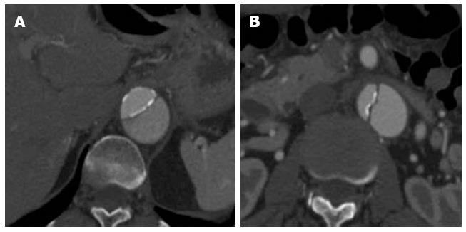
Figure 5 A 59-year-old lady with a type B aortic dissection.
Axial images from a computed tomography aortogram show atherosclerotic calcifications outlining the true lumen at the lower thoracic aorta (A). The true lumen is of smaller calibre and shows early and more intense enhancement than the false lumen at this level. More inferiorly at the level of the left renal vein (B), eccentric intimomedial flap calcification (note the calcification along the true luminal aspect of the flap) is exquisitely demonstrated.
- Citation: Hallinan JTPD, Anil G. Multi-detector computed tomography in the diagnosis and management of acute aortic syndromes. World J Radiol 2014; 6(6): 355-365
- URL: https://www.wjgnet.com/1949-8470/full/v6/i6/355.htm
- DOI: https://dx.doi.org/10.4329/wjr.v6.i6.355


