Copyright
©2014 Baishideng Publishing Group Inc.
World J Radiol. Jun 28, 2014; 6(6): 329-343
Published online Jun 28, 2014. doi: 10.4329/wjr.v6.i6.329
Published online Jun 28, 2014. doi: 10.4329/wjr.v6.i6.329
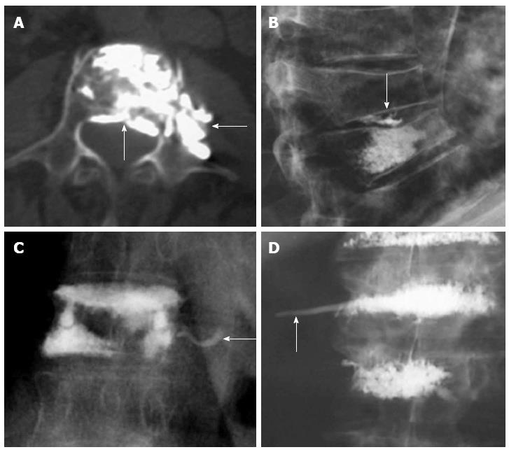
Figure 8 Axial computed tomography shows extravasations to epidural space and paravertebral area (arrows) (A), leakage to the intervertebral disc (arrow) (B), venous leakage (arrow) (C), tail of cement in the path of the needle (arrow) (D).
- Citation: Santiago FR, Chinchilla AS, Álvarez LG, Abela ALP, García MDMC, López MP. Comparative review of vertebroplasty and kyphoplasty. World J Radiol 2014; 6(6): 329-343
- URL: https://www.wjgnet.com/1949-8470/full/v6/i6/329.htm
- DOI: https://dx.doi.org/10.4329/wjr.v6.i6.329


