Copyright
©2014 Baishideng Publishing Group Inc.
World J Radiol. Jun 28, 2014; 6(6): 313-328
Published online Jun 28, 2014. doi: 10.4329/wjr.v6.i6.313
Published online Jun 28, 2014. doi: 10.4329/wjr.v6.i6.313
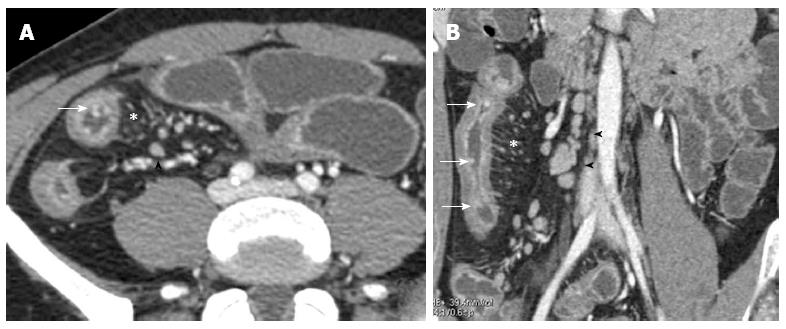
Figure 8 Transverse (A) and coronal (B) computed tomography show bowel wall thickening and mucosal hyper-enhancement with pseudo polyps (white arrows) as well as mesenteric lymph nodes, that are irregular in size and shape (black arrowhead) and increased mesenteric vascularity (asterisk).
These features may be detected in case of acute exacerbation on a background of longstanding disease.
- Citation: Casciani E, Vincentiis CD, Polettini E, Masselli G, Nardo GD, Civitelli F, Cucchiara S, Gualdi GF. Imaging of the small bowel: Crohn’s disease in paediatric patients. World J Radiol 2014; 6(6): 313-328
- URL: https://www.wjgnet.com/1949-8470/full/v6/i6/313.htm
- DOI: https://dx.doi.org/10.4329/wjr.v6.i6.313


