Copyright
©2014 Baishideng Publishing Group Inc.
World J Radiol. Jun 28, 2014; 6(6): 274-283
Published online Jun 28, 2014. doi: 10.4329/wjr.v6.i6.274
Published online Jun 28, 2014. doi: 10.4329/wjr.v6.i6.274
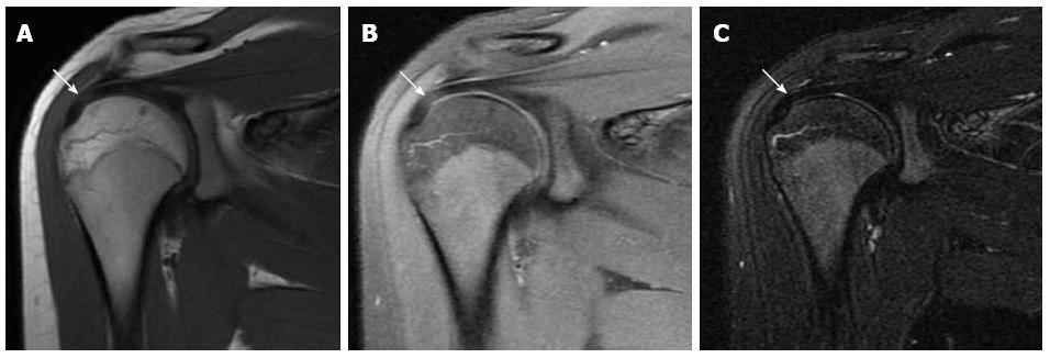
Figure 9 Magic angle artifact.
A: Coronal oblique T1-weighted image. B: Coronal oblique fat-sat proton-density image. Images (A) and (B) show focal high signal intensity within the distal supraspinatus tendon (arrow); C: Coronal oblique fat-sat T2-weighted image shows normal signal of the supraspinatus tendon (arrow). Final diagnosis was magic angle artifact with normal tendon.
- Citation: Tawfik AM, El-Morsy A, Badran MA. Rotator cuff disorders: How to write a surgically relevant magnetic resonance imaging report? World J Radiol 2014; 6(6): 274-283
- URL: https://www.wjgnet.com/1949-8470/full/v6/i6/274.htm
- DOI: https://dx.doi.org/10.4329/wjr.v6.i6.274


